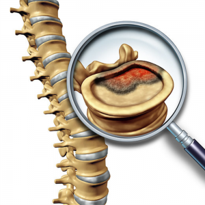
Spinal Tumors
A spinal tumor is an abnormal growth that develops in the spinal canal or within the bones of the spine. Spinal tumors may be primary or secondary. Primary tumors originate in the spine or spinal cord, and secondary tumors (metastasis) result from cancer spreading from another site to the spine. Spine tumors may grow in any area of the spine, from cervical to sacrum.
Causes of Spinal Tumors
It’s not clear why most spinal tumors develop. Some of them may be attributed to exposure to cancer-causing agents. Spinal cord lymphomas, which are cancers that affect lymphocytes (a type of immune cell), are more common in people with compromised immune systems. In some cases, spinal cord tumors are linked to known inherited syndromes, such as neurofibromatosis 2 and von Hippel-Lindau disease.
Types of Spinal Tumors
Spinal tumors may be benign or malignant. They are classified into 3 major groups: intramedullary, intradural-extramedullary and vertebral column tumors.
Vertebral column tumors
These tumors involve the bones of the vertebral column. The majority of these tumors are metastatic. That is, the original tumor developed in another organ and has spread to the vertebral column, usually through the bloodstream. The most common metastatic spinal tumors in women are from the breast and lung. In men, metastatic spinal tumors are most often from the prostate and lung.
Tumors arising from vertebral bone and cartilage cells also occur in the spine, although less frequently. Examples of these primary spinal column tumors include:
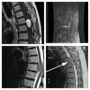
Figure 1. Different spinal tumors observed on MRI
Benign
- Osteoid osteoma
- Osteoblastoma
- Giant cell tumor
Malignant
- Osteogenic sarcoma
- Chordoma
- Chondrosarcoma
- Ewing’s sarcoma
- Metastasis (breast, lung, prostate, lymphoma, kidney)
Intradural-extramedullary tumors
Intradural-extramedullary tumors grow within the spinal canal. These tumors are located inside the dura, but outside the spinal cord. Meningiomas and nerve sheath tumors (schwannomas and neurofibromas) comprise the majority of this subset of spinal tumors.
- Meningiomas arise from the dura mater, the thin membrane that surrounds the spinal fluid and spinal cord. Meningiomas are most common in middle-aged and older women. These tumors are usually benign.
- Schwannomas and neurofibromas arise from the nerve roots that leave the spinal cord. Like meningiomas, nerve sheath tumors are usually benign.
- Filum terminale ependymomas occur just below the spinal cord, in the lumbar spinal canal. Nearly all are benign. These tumors may be large and may adhere to many nerves, making total removal sometimes difficult.
Intramedullary tumors
Intramedullary tumors are located inside the substance of the spinal cord.
These tumors usually arise from glia (supporting cells) within the spinal cord. Astrocytomas and ependymomas account for the majority and occur with about equal frequency, although astrocytomas are more common in children and ependymomas more common in adults.
Hemangioblastomas, tumors of blood vessels, are less frequent and sometimes occur in conjunction with Von Hippel Lindau disease. Intramedullary tumors occur most often in the cervical spinal cord, or spinal cord in the neck, and are usually benign.
Symtoms of Spinal Tumors
Depending on size and location, spinal cord tumors can cause different signs and symptoms. Spinal tumors progress at different rates depending on the type of tumor.
Signs and symptoms may include:
- Pain at the site of the tumor due to tumor growth
- Back pain, often radiating to other parts of your body
- Feeling less sensitive to pain, heat, and cold
- Loss of bowel or bladder function
- Scoliosis
- Difficulty walking, sometimes leading to falls
- Back pain that’s worse at night
- Loss of sensation or muscle weakness, especially in your arms or legs
- Muscle weakness, which may be mild or severe, in different parts of your body
Diagnosis of Spinal Tumors
A thorough medical examination with emphasis on back pain and neurological deficits is the first step to diagnosing a spinal tumor. Radiological tests are required for an accurate and positive diagnosis. The most useful diagnostic test is the magnetic resonance (Fig 1).
Treatment of Spinal Tumors
Treatment of spinal tumors is often multidisciplinary, incorporating the expertise of spinal surgeons, medical oncologists, radiation oncologists, and other medical specialists. Tumors that are asymptomatic or mildly symptomatic and do not appear to be changing or progressing may be observed and monitored with regular MRIs. Some tumors respond well to chemotherapy and others to radiation therapy.
Indications for surgery
This is often the treatment of choice for tumors that can be removed with an acceptable risk of spinal cord or nerve injury damage. Newer techniques and instruments allow neurosurgeons to reach tumors that were once considered inaccessible. The high-powered microscopes used in microsurgery make it easier to distinguish tumor from healthy tissue. But even with the latest technological advances in surgery, not all tumors can be removed entirely. When the tumor can’t be removed completely, surgery may be followed by radiation therapy or chemotherapy or both.
Awesome Doctors for your Neurosurgery Needs

Dr. JJ Ramirez
Neurocirujano
Dr. Ramirez attended medical school at the Universidad Autonoma of Guadalajara School of Medicine in Guadalajara Mexico. Dr. Ramirez performed his Neurosurgery training at the National Institute of Neurology and Neurosurgery in Mexico City

Dr. Ramiro Pérez
Neurosurgeon
Dr. Ramiro Pérez attended medical school at the University of Guadalajara School of Medicine in Guadalajara México. Dr. Pérez performed his Neurosurgery training in Centro Medico Siglo XXI IMSS and has been practicing his specialty for more than 13 years.

Dr. Felipe Nares
Neurosurgeon
Dr. Nares attended medical school at the University of Aguascalientes, He is trained to perform anterior and lateral approaches to the spine from the cervical to the lumbar spine, as well as minimally invasive surgery.

Dr. Luis A. Robles
Neurosurgeon
Dr. Luis Robles has been practicing neurosurgery specialty for 20 years. Dr. Robles is academically active, he participates as a section editor in the World Neurosurgery journal and he has published several articles in different neurosurgery international journals.